3, Roles of dying cells as a signal center
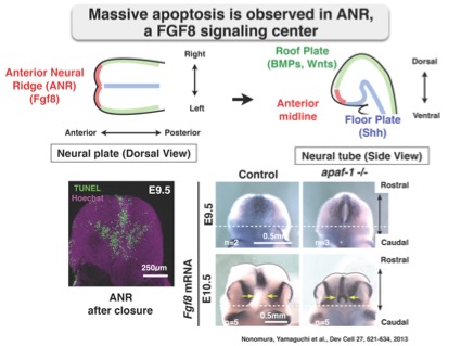
Apoptotic cells are abundantly observed in the early developing brain. It was proposed that apoptosis-deficiency causes brain overgrowth, but we demonstrated that brain malformations in apoptosis-deficient mutants are due to insufficient brain ventricle expansion as a result of uncompleted cranial neural tube closure. Apoptosis eliminates Fgf8-expressing cells in the anterior neural ridge (ANR), which acts as an organizing center of the forebrain by producing FGF8 morphogen. Deficiency of apoptosis leads to the accumulation of non-proliferative undead cells in the ventral part of the ANR. The undead cells in apoptosis-deficient mutants express Fgf8 continuously, which perturbs gene expression in the ventral forebrain. From these observations, we proposed that apoptosis within a specific subdomain of the ANR is required for correct temporal elimination of FGF8-producing region within a limited developmental time window, thereby ensuring proper forebrain development.
Cell death is thought to be widely observed during development, tissue homeostasis and disease condition. However, reality is different. Even in the sensitive TUNEL assay, detection of apoptosis is not easy. Apoptotic cells are rapidly removed from body. We can only recognize the area massive cell death occurs. Live cell imaging using fluorescence resonance energy transfer (FRET) technology permits us to monitor cell-signaling activities simultaneously with cell behavior in real time. Live FRET imaging with a fast-scanning confocal microscope enable us to monitor in vivo dynamics of caspase activation. SCAT (sensor for caspase activation based on FRET) consists of ECFP and Venus, both of which are connected with a linker sequence containing cleavage sites of caspase. We succeeded in monitoring caspase-3 activation by SCAT3 in living Drosophila pupa and mouse embryo.
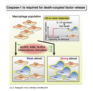
We have generated FRET sensor for activated caspase-1, named SCAT1. We examined the spatial and temporal activation of caspase-1 in macrophages, and found that activation of caspase-1 was the manner of all-or-none (digital) at the single-cell level, with similar activation kinetics irrespective of the type of inflammasome or the intensity of the stimulus. Caspase-1 activated macrophage die immediately and real-time concurrent detection of caspase-1 activation and IL-1b release demonstrated that dead macrophages release a local burst of IL-1b in a digital manner. These dying macrophages as the main source of IL-1b within cell populations. These results highlight the value of single-cell analysis in understanding of the inflammasome system and chronic inflammatory diseases. Thus combination of live imaging and genetics revealed the active roles of caspase-mediated cell death during development and inflammation.
References
Liu, T., Yamaguchi, Y., Shirasaki, Y., Shikada, K., Yamagishi, M., Hoshino, K., Kaisho, T., Takemoto, K., Suzuki, T., Kuranaga, E., Ohara, O., and Miura, M.: Single-cell imaging of caspase-1 dynamics reveals an all-or-none inflammasome signaling response. Cell Rep 8, 974-982, 2014.
Yamaguchi, Y., Kuranaga, E., Nakajima, Y., Koto, A., Takemoto, K., and Miura, M.: In vivo monitoring of caspase activation using a fluorescence resonance energy transfer-based fluorescent probe. Regulated Cell Death. ed. Ashkenazi, A., Wells , J., Yuan, J. Methods Enzymol. 544, 299-325, 2014
Nonomura, K., Yamaguchi, Y., Hamachi, M., Koike, M., Uchiyama, Y., Nakazato, K., Mochizuki, A., Sakaue-Sawano, A., Miyawaki, A., Yoshida, H., Kuida, K., and Miura, M.: Local apoptosis modulates early mammalian brain development through the elimination of morphogen producing cells. Dev. Cell 27, 621-634, 2013
Yamaguchi, Y., Shinotsuka, N., Nonomura, K., Takemoto, K., Kuida, K., Yoshida, H., and Miura, M.: Live imaging of apoptosis in a novel transgenic mouse highlights its role in neural tube closure. J. Cell Biol. 195, 1047-1060, 2011
Back to Research page
---------------------------------------------------------------------------------------------------------------------
This page is administered by Department of Genetics All Rights Reserved, Copyright © 2019 Department of Genetics
© 2010 IDEN-Homepage